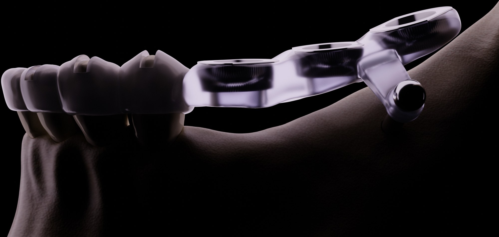
A decade ago, a "difficult canal" meant longer chair time, more radiographs, and a fair amount of clinical intuition. Today, that same case can be planned in three dimensions before the handpiece ever touches the tooth. CBCT imaging combined with 3D treatment planning software has quietly become one of the most consequential shifts in endodontics, and clinics across the UAE are beginning to treat it not as a luxury add-on, but as a genuine diagnostic standard for complex cases.
What makes this shift significant isn't just sharper images. It's the fact that a single CBCT scan can now feed directly into diagnosis, treatment planning, patient communication, and even guided access template design, all from one dataset. Understanding what this technology actually adds, and where its limits are, matters for any clinician deciding whether and when to bring it into their diagnostic workflow.
Why 2D Imaging Was Never Enough
A conventional periapical radiograph compresses a three-dimensional root canal system into a flat, two-dimensional image. Extra canals, isthmuses, and curvatures in the buccolingual plane are frequently invisible until the clinician is already inside the tooth, because the radiograph simply cannot show depth in that direction.
This limitation isn't a minor inconvenience. Studies on root canal anatomy have repeatedly shown that a meaningful proportion of teeth, particularly mandibular molars and maxillary premolars, have canal configurations that are easy to misread or entirely miss on 2D imaging alone. A second mesiobuccal canal in a maxillary molar, for example, is a classic case where 2D radiography routinely underestimates what is actually there.
CBCT removes that blind spot entirely, giving a true 3D view of root canal anatomy, periapical pathology, and proximity to critical structures such as the inferior alveolar nerve or maxillary sinus, viewed from any angle the clinician chooses.
What a CBCT Scan Actually Captures
Modern CBCT units allow clinicians to select a field of view appropriate to the case, from a small, high-resolution scan covering a single tooth or quadrant, to a larger field of view for full-arch assessment. Smaller fields of view generally offer higher resolution and lower radiation dose, making them well suited to focused endodontic diagnosis rather than routine full-mouth screening.
The resulting dataset can be viewed slice by slice in any plane — axial, sagittal, or coronal — giving a level of anatomical detail that simply isn't available from a flat image. This is particularly valuable for assessing root curvature in the buccolingual direction, which a periapical radiograph cannot show at all.
From Diagnosis to Design: The 3D Planning Workflow
Once a CBCT scan is captured, treatment planning software allows the clinician to visualise the entire canal system from any angle, measure canal curvature and length with precision, and identify calcifications long before opening the tooth. This same dataset can then be used to design a 3D-printed guided access template, connecting diagnosis directly to execution in a single digital workflow.
Beyond access planning, the same scan supports a range of diagnostic tasks: distinguishing a true periapical lesion from anatomical structures that mimic one on a 2D film, assessing the extent of external or internal resorption, evaluating suspected vertical root fractures, and planning retreatment cases where the original canal path has become obscured by previous restorative or endodontic work.
- Earlier, accurate detection of missed or accessory canals
- Reduced radiation exposure per scan compared to older CBCT generations, especially with small field-of-view protocols
- Direct integration with guided access template design
- More reliable assessment of periapical pathology, resorption, and suspected fractures
- Better case documentation for referrals, insurance, and patient communication
The tooth does not change. What changes is how much of its true anatomy we are willing to see before we start drilling.
Radiation, Cost, and When It's Actually Indicated
CBCT is not intended to replace routine periapical radiography for every root canal case, and using it indiscriminately adds unnecessary radiation exposure and cost without meaningful clinical benefit. The principle that should guide CBCT usage is the same one that governs all diagnostic imaging: justified use, keeping exposure as low as reasonably achievable for the clinical question being asked.
In practice, this means CBCT tends to be most valuable for retreatments, teeth with calcified or unusually curved canals, suspected extra roots or canals, unresolved periapical pathology after seemingly adequate treatment, trauma cases, and situations where a guided access template is being planned.
What This Means for Everyday Practice
Clinics that have integrated CBCT into their diagnostic protocol, and used it selectively rather than universally, consistently report fewer surprises mid-treatment and more informed conversations with patients about complexity and prognosis before treatment even begins. It also strengthens referral letters and case documentation considerably, since a 3D dataset communicates far more to a receiving specialist than a written description alone.
The Skill Behind the Scan
Owning or accessing a CBCT machine is only the first step. Interpreting the scan accurately, distinguishing genuine pathology from anatomical variation, and translating findings into a treatment plan or a guided access template, requires focused training. This is precisely the gap that ACAD's guided endodontics workshop is built to close, taking clinicians from CBCT interpretation through to hands-on guided access preparation.
Learn the Complete CBCT-to-Guide Workflow
ACAD's hands-on Guided Endodontics workshop covers CBCT interpretation, digital guide design, and chairside execution. CPD Standards Office UK accredited.